Comprehensive Treatment
Except for the few specific situations in which interceptive treatment can fully correct a crowding or bite problem, the majority of malocclusions require comprehehensive treatment to fully align the teeth and correct the bite.
Comprehensive treatment can be broadly divided into 3 categories:
1. Braces only
2. Braces in combination with a plate or expander (“two phase” treatment)
3. Braces in combination with jaw surgery
Braces Only
Case 1
Class I crowding (Jaw relation normal, teeth crowded)

After treatment with full upper and lower braces. Two premolar teeth extracted from the top jaw for space.

Case 2
Class III crowding (Bottom jaw overshot and tooth crowding)

Treated with full upper and lower braces. Two premolar teeth extracted from the bottom jaw to compensate for the overshot bite. Top jaw treated by expansion to make space and no tooth extractions.

Case 3
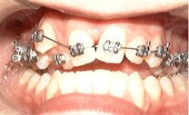
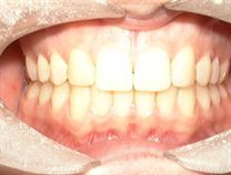
Class I missing upper teeth
Upper permanent lateral incisors congenitally absent (tooth germ never formed).
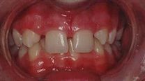
Treated with full upper and lower braces to align upper teeth and open deep bite. Later upper canine teeth reshaped to look like the missing lateral incisors.

Case 4
Impacted canine teeth
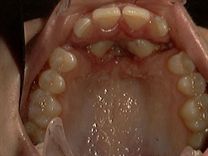
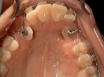
The teeth maybe impacted in the roof of the mouth or less commonly, between the adjacent teeth. The impacted canine teeth are uncovered first – this would be done under local or general anaesthetic, depending on the depth of the impaction. The braces are generally placed about 2 weeks later, giving time for the gum to heal around the exposed tooth.
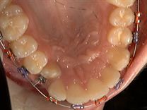
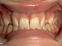
Photos: Single tooth exposed in roof of mouth, Radiograph shows the impacted , canine tooth on the upper right side, Both canines exposed and aligned from roof of mouth, No sign of upper canine teeth radiograph shows both upper canine teeth in roof of mouth, canine teeth uncovered, Aligning with braces, Both canines impacted between adjacent teeth - Canine teeth exposed and aligned with braces.


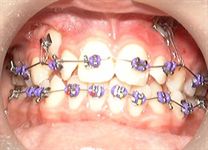
Case 5
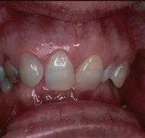
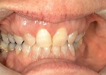
Deep bite (Class II division 2 malocclusion)
Causing damage to the gum on the bottom teeth. Full braces were required to open the bite and align the teeth accordingly. If left untreated, the lower gum would have been stripped away to a point where the bottom front teeth could be lost.
Photos: Deep Bite, Bite Opened
Two phase treatment
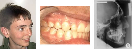
Case 1
Class II upper incisor protrusion (“Buck teeth” and undershot bottom jaw)
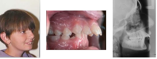
Child treated with a functional plate first to correct the incisor protrusion, followed by upper and lower braces to fully align the teeth and “sock in” the bite. Tooth extractions were not required.
Braces combined with jaw surgery
Jaw surgery is generally recommended by orthodontists when the discrepancy in the jaw relationship is too severe to compensate with braces treatment alone and in adult patients with undershot jaws, where there is no growth left to help correct the incisor protrusion. Jaw surgery is generally not carried out until growth is substantially complete.
Some jaw discrepancies requiring surgery for correction:
1. Undershot bottom jaw in an adult
2. Overshot bottom jaw/ underdeveloped top jaw
3. Open bite
4. Excessively narrow top jaw in an adult, requiring expansion
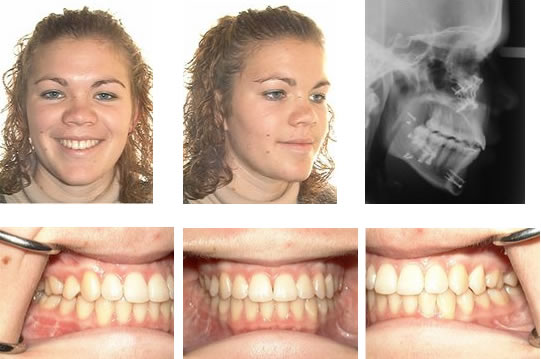
Case 1
Overshot bottom jaw

After treatment with braces and jaw surgery
Undershot bottom jaw
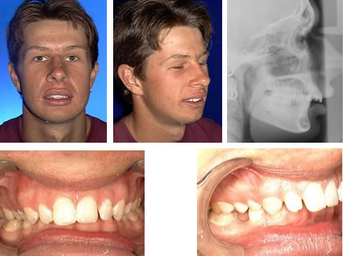
Braces to align teeth prior to surgery
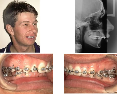
After surgery
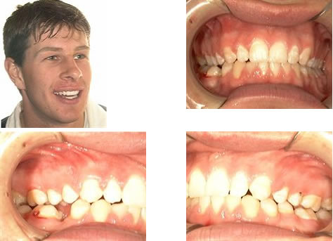
Underdeveloped top jaw

After surgery

Surgery to assist expansion of the top jaw in adult patient
The top jaw forms as two halves, which are joined together and to other facial bone by sutures. In adult patients, the sutures of the top jaw are fused, unlike in a child. So, before the top jaw can be expanded in an adult, the sutures need to be surgically split, allowing the two halves of the jaw to be separated. Once the expansion is done, the jaw bone heals back as a single unit again.
Before surgery, with RME appliance in place but not activated, on right, after surgery with appliance fully activated. Note gap between incisor teeth

RME is left in place for 4 weeks after to bone healing in jaw. Note gap between incisors has closed spontaneously.
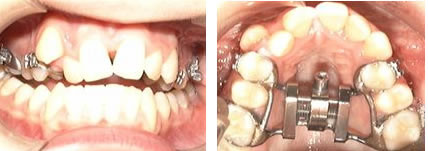
Braces placed to start aligning the top teeth. Bottom braces will be placed later.
